Endometrial Cancer
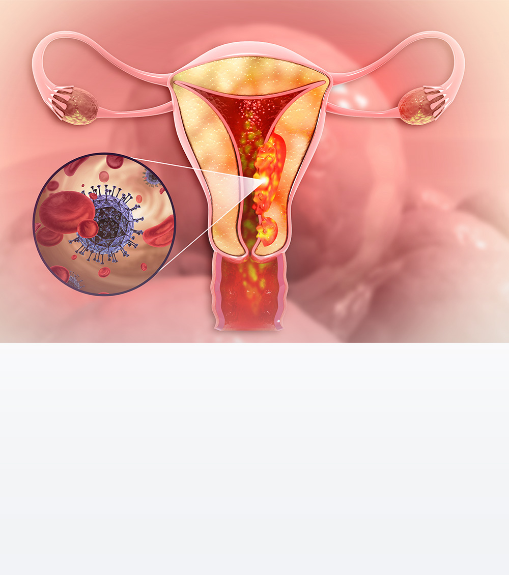
The uterus has three layers which include the outer perimetrium (serosa), the middle one is myometrium and the innermost lining is the endometrium. Monthly changes in endometrium result in menstrual cycles in women.
The two types of cancer that occur in the uterus are endometrial carcinoma, which is more common and forms in the endometrium and the second is sarcoma which stems from uterine myometrium.
The uterus is a hollow and pear-shaped organ situated in the pelvic region and part of the female reproductive system where fetal growth takes place. The endometrium is the innermost lining of the uterus and tumours that develop in this innermost lining are known as endometrial cancer or uterine cancer.
Symptoms of Endometrial Cancer:
The most common symptom of endometrial cancer is abnormal vaginal bleeding. It can be
Types of Endometrial Cancer
There are two types of endometrial carcinoma:
- Type I or endometroid adenocarcinoma is associated with excess estrogen secretion less aggressive type and spread relatively slowly.
- Type II or serous carcinoma or clear cell carcinoma are non-estrogen dependant and aggressive types of cancer.
Adenocarcinoma, Uterine carcinosarcoma, Squamous cell carcinoma, Small cell carcinoma, Transitional carcinoma, and Serous carcinoma are multiple forms of endometrial cancers based on histopathological studies and Clear cell carcinoma, Mucinous adenocarcinoma, Serous adenocarcinoma, etc., are some of the less common types of cancer.
Based on pathological studies endometrial cancers are divided into grades 1, 2 and 3 depending on the cell structure, which is crucial to plan treatment strategies.

Diagnostics Facilities
Advanced Cancer Diagnostics
Advanced Cancer Treatment
General Diagnostic Facilities
Causes of Endometrial Cancer:
Endometrium growth varies according to the levels of estrogen and progesterone hormones secreted from ovaries. Excess levels of estrogen are responsible for the proliferation of tumours in the endometrium, causing Type I and Type II endometrial cancers.
Non-modifiable
- Age: Chances of contracting cancer increase as we age.
- Family history: About 5-10% of endometrium cancers are related to family history. History of multiple family members having colon cancer increases the possibility of familial inheritance and should be ruled out with genetic testing; Lynch syndrome is a condition where the chances of multiple family members having various cancers are high.
- Early menarche and late menopause increase the duration of menstruation in years, exposing the endometrium to the estrogen hormone for a longer period, which increases the chances of endometrial cancer.
- Estrogen-secreting ovarian cancers can also cause endometrial cancer.

Modifiable causes
Most of these causes are related to the secretion of excess Estrogen due to external causes.
- Obesity: Once women pass the menstruation phase the ovarian function stops but estrogen hormone can be produced from fat cells known as adipose tissue of the body. This is one of the most common causes of endometrium cancer. Obesity, diabetes and high blood pressure usually coexist in patients and are known as the triple syndrome of endometrial cancer and are responsible for excessive estrogen production in the body.
- Diet and physical activity: High-fat content in diets and sedentary lifestyles are related to increased body weight and in turn are related to endometrial cancer.
- Polycystic ovarian syndrome: This condition is associated with the secretion of excess estrogen and androgen hormones causing endometrial cancer.
- Taking hormones, known as hormone replacement therapy to combat postmenopausal symptoms, that contain Estrogen alone but not progesterone also leads to endometrial cancer.
- Hormone therapy for breast cancer: Tamoxifen is an anti-cancer drug used for the treatment of breast cancer and can cause endometrial hyperplasia (increase in thickness of endometrial lining). The association of tamoxifen with endometrial cancer has been proven since the chances are not very high and considering the risk-benefit ratio tamoxifen is considered an essential medicine for breast cancer hence the use of same should not be stopped.
Prevention of Endometrial Cancer
With no defined screening protocol for endometrial cancer awareness is the best way to prevent these cancers and any abnormal uterine bleeding, especially post-menopausal bleeding should not be ignored.
It is best to adopt the following strategies to reduce the risk of endometrial cancer:

Investigations to diagnose Endometrial Cancer
Physical examination of the body to detect the presence of lumps, lymph nodes or any unusual signs of disease along with a detailed analysis of the patients’ health past illnesses and treatments is crucial to determine the presence of endometrial cancer
Imaging Ultrasonogram (USG): Transabdominal ultrasonogram of the abdomen and pelvis is a safe and simple way of detecting any abnormality in the uterus and ovaries, based on USG findings a biopsy is performed.
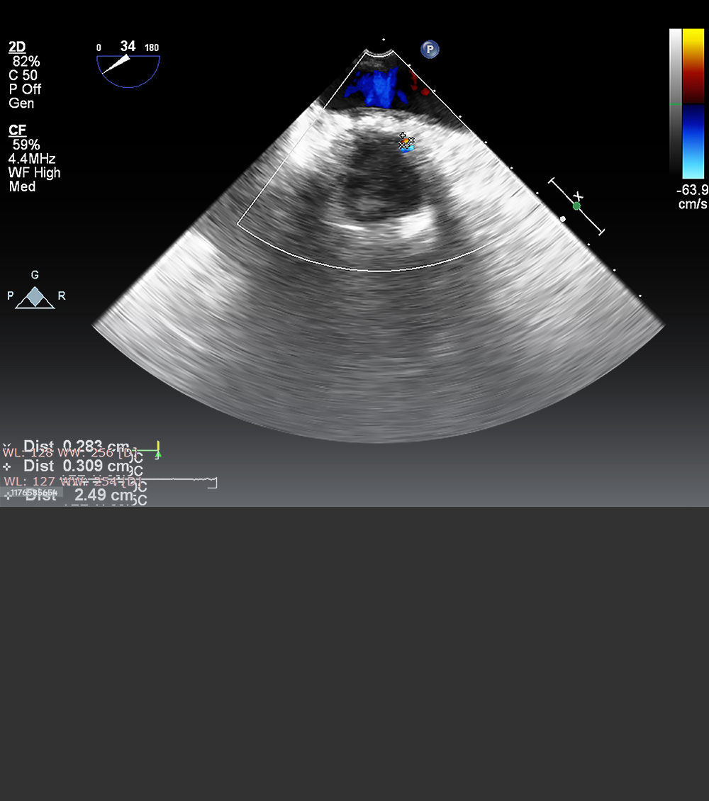
Transvaginal ultrasound: In TVS or transvaginal ultrasound, an ultrasound transducer or a probe-like structure is inserted into the vagina which sends high-energy sound waves to the internal tissues and organs like the vagina, uterus, fallopian tubes and bladder.
CT scan: After confirming the diagnosis with biopsy a contrast-enhanced CT scan of the abdomen is performed to gain more detailed information on the tumour.
MRI: MRI is performed on diagnosing the presence of cancer cells in the pelvic region to know about the spread of the disease, which helps in making relevant treatment plans.
PETCT Scan: If initial investigations indicate an advanced stage or recurrence of the disease in people who already if somebody already had endometrial cancer a PETCT scan is done to understand the spread of the disease.
Biopsy: Biopsy is the process of collecting cell/tissue samples from the inner lining of the uterus and is performed to detect abnormal uterine bleeding and is studied under the microscope by pathologists to check for the presence of abnormal tissues. A PAP smear commonly done for cervical cancer screening is not helpful because endometrial cancer forms inside the uterus and samples of endometrial tissue should be taken and checked under the microscope to detect the presence of cancer cells.
The following procedures are used to diagnose these cancers.
Endometrialpipette biopsy: This clinical procedure does not require any type of anaesthesia, since it is painless and just causes mild discomfort. A thin and flexible tube is inserted through the cervix into the uterus to take tissue samples from the endometrium.
- Dilatation and curettage: The D&C procedure requires mild anaesthesia, wherein, the cervix is dilated and a spoon-shaped instrument is inserted into the uterus to collect tissue samples which are then studied under the microscope to detect the presence of cancer cells.
- Hysteroscopy: This procedure is done under the influence of local anaesthesia, wherein a hysteroscope is used to examine the uterus linings to detect the presence of abnormal tissues. It requires anaesthesia.The hysteroscope is a thin tube-like instrument with a light source and lens with a tool to collect tissue samples. It is similar to the D&C procedure but offers the consultant a clear view of the inner linings of the uterus.

Treatment for Endometrial Cancer
The treatment for endometrial cancer varies with the stage of cancer, age and fertility status endometrial carcinoma is treated. Modern science offers possibilities for fertility preservation in younger women who wish to have children after cancer treatments.
For older women who have had children past their childbearing age the following treatment options are available:
Surgery:
Surgical procedures are planned based on the patient’s age and cancer staging and involve removal of the uterus, both ovaries, along with fallopian tubes and lymph nodes. Both laparoscopic (minimally invasive) and open surgeries can be performed with the former delivering the best outcomes with less pain and speedy recovery.
The specimen is sent for a detailed histopathological examination which aids in planning further treatment strategies.
Radiation therapy:
- Based on histopathology findings, the need for radiation therapy is determined, and early-stage cancers do not require further treatment. Brachytherapy is an internal radiation therapy used for cancers spread deep inside the uterus.
- External beam radiation therapy (EBRT) is used to treat tumours, which has spread to the lymph nodes.
Chemotherapy:
The need for chemotherapy is determined based on histopathological findings and is given to patients to avoid further spread of tumours.
Hormone therapy:
Endometrium cancer is hormone-sensitive cancer and the hormone therapy approach, in the form of intrauterine implants, is adopted when other treatment options are not possible due to various reasons.
Doctors

Dr. Deepti Mishra

Dr. Bhavesh Poladia


